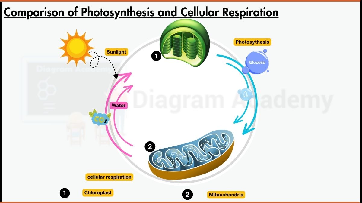
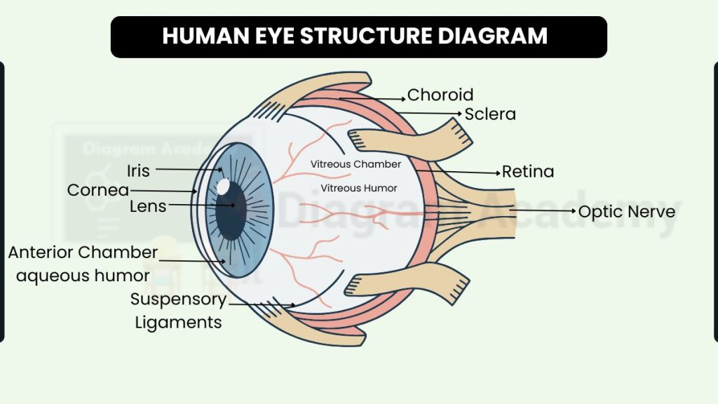
Labelled Diagram of Human Eye structure
The human eye is a remarkable sensory organ that works like a natural camera, helping us see, differentiate millions of colors, and stay connected to our surroundings. The human eye structure diagram shows both the external and internal parts of the eye, each playing an important role in vision. Externally, parts like the sclera, cornea, iris, and pupil help protect the eye and control how much light enters. Internally, structures such as the lens, retina, optic nerve, aqueous humour, and vitreous humour work together to focus light, form images, and send signals to the brain. Understanding the eye diagram helps students learn how vision works and why each part is essential for clear sight.
FAQs – Human Eye Structure Diagram
Q1. What is shown in the human eye structure diagram?
It shows all the major parts of the eye—both external and internal—and how they work together for vision.
Q2. What are the external parts of the eye?
Sclera, conjunctiva, cornea, iris, and pupil.
Q3. What does the lens do?
The lens bends (refracts) light so it focuses clearly on the retina.
Q4. What is the function of the retina?
The retina captures the image and sends signals to the brain through the optic nerve.
Q5. Why is the cornea important?
It protects the eye and helps focus light entering the eye.
Q6. What is the difference between aqueous humour and vitreous humour?
Aqueous humour is the watery fluid in front of the lens, while vitreous humour is the gel-like fluid behind the lens.
Q7. How does the iris help us see?
It controls the size of the pupil and regulates how much light enters the eye.