What Does a Gram Bacteria Diagram Show?
A gram diagram visually compares the structural differences between Gram-positive and Gram-negative bacteria. These diagrams focus mainly on the cell wall layers, which determine how bacteria respond to Gram staining.
The diagram of gram positive and gram negative bacteria helps students clearly see structural differences that are difficult to understand through text alone.
Diagram of Gram-Positive Bacteria
In a diagram of gram positive bacteria, you will notice:
- A thick peptidoglycan layer
- No outer membrane
- Teichoic acids embedded in the cell wall
- Single cell membrane beneath the wall
The diagram of gram positive cell wall highlights that the peptidoglycan layer is very thick, which allows these bacteria to retain the purple stain during Gram staining.
Key Visual Features:
- Thick outer wall
- One membrane
- No lipopolysaccharide (LPS) layer
Diagram of Gram-Positive Bacterium
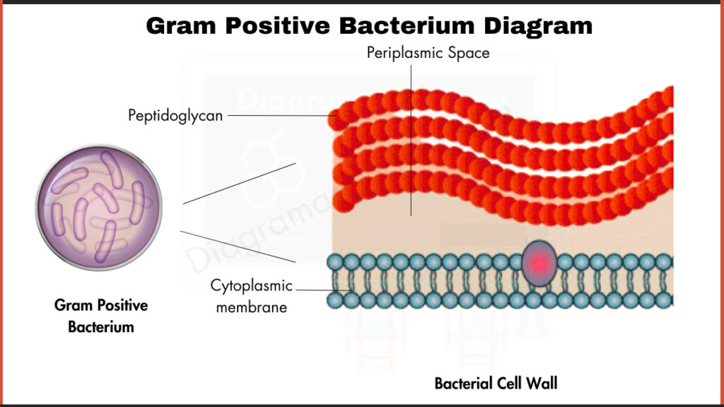
Diagram of Gram-Negative Bacteria
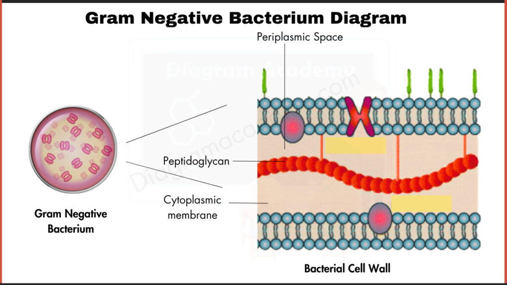
The diagram of gram negative bacteria looks different because these bacteria have:
- A thin peptidoglycan layer
- An outer membrane
- Lipopolysaccharide (LPS) layer
- Two membranes (inner and outer)
In the gram positive and gram negative bacteria diagram, Gram-negative bacteria appear more complex due to the additional outer membrane.
Key Visual Features:
- Thin cell wall
- Two membranes
- Periplasmic space
- LPS layer
Side-by-Side Comparison Diagram
A diagram of gram positive and negative bacteria typically shows:
| Feature | Gram-Positive | Gram-Negative |
| Peptidoglycan Layer | Thick | Thin |
| Outer Membrane | Absent | Present |
| LPS Layer | No | Yes |
| Stain Color | Purple | Pink/Red |
This type of gram positive and gram negative diagram is commonly used in biology textbooks and labmanuals.
FAQs Frequently Asked
Here are the answer to common question about Gram-Positive and Gram-Negative Bacteria.
Q1: What is shown in a gram diagram?
It shows the structural differences in the cell walls of Gram-positive and Gram-negative bacteria.
Q2: Why is the gram positive cell wall thicker?
It contains a thick peptidoglycan layer.
Q3: What makes Gram-negative bacteria different in diagrams?
They have an additional outer membrane and an LPS layer.
Q4: Why are these diagrams important in microbiology?
They explain staining results and antibiotic sensitivity differences.